
AMERICAN ACADEMY OF ARTIFICIAL INTELLIGENCE IN DENTISTRY®
The AAAI-D Brief
October 2026
The Academy’s October review of published research covers an evaluation of AI caries detection that found high specificity and lower sensitivity for dentin lesions; a pooled analysis of periodontal AI covering more than 50,000 radiographs; imaging AI that performed better when patient data accompanied the image; an analysis of the chatbots patients consult for health information; a study of how the presentation of AI advice affects clinicians; a neural network for an orthodontic treatment decision, tested on external data; and a university program in which dental students help train an AI system.
This edition is the Academy’s October update.
This issue presents the Academy’s review of research published this month, together with shorter reports and Academy news.
CONTENTS
FEATURED STUDIES
A Reality Check on AI Caries Detection
The Chart Helps the Algorithm Read the Film
Periodontitis AI, Pooled: Strong Scores, and Proof Still to Come
When Patients Ask the Chatbot First
Surgery First, or Orthodontics First? A Neural Network Weighs In — Then Faces Outside Data
Same AI Advice, Different Dress: How Presentation Changes What Clinicians Do
ALSO WORTH KNOWING
AI Finds the Impacted Canine — and Misses the Resorption
Pulp Stones Are Easy to Find — Until the Tooth Has Been Restored
Timing Expansion: AI Reads the Palatal Suture — on a Scanner It Never Trained On
A General-Purpose Chatbot, Tested on Oral Lesions
Measuring “Black Triangles” From a Photograph
One Model, 27 Structures: Automated CBCT Anatomy
Featured Studies
The month’s most substantial studies, in full.
A Reality Check on AI Caries Detection
In a study of 4,000 first permanent molars in children aged 6–12, published in BMC Oral Health, an AI-assisted detection system working from 1,000 panoramic radiographs was highly specific (99.6%) but identified only about one in three dentin lesions at the threshold examiners used (sensitivity 32.1% for dentin caries at the RA3+ threshold, against ICCMS criteria; agreement with examiner consensus was fair, κ = 0.389). Performance improved substantially for advanced lesions, reaching 80.2% at RC5 and 93.4% at RC6; overall accuracy was 78.9%. The disease burden in this sample was not small: at the child level, 56.8% of the children had at least one advanced (RA3+) lesion.1
The authors conclude that the system should serve as an adjunct to clinician-led assessment, not as a standalone screening or triage tool.1
The pattern is mixed: very few false alarms, low sensitivity for dentin lesions at the threshold examiners used, and much higher detection of advanced lesions.
The Chart Helps the Algorithm Read the Film
Most dental imaging AI looks at the image alone. A study in BMC Medical Imaging combined panoramic radiographs with structured patient data to grade periodontal bone-resorption severity across 988 radiographs, and the combined model — a DenseNet-121 image network joined to the chart data — outperformed both image-only and data-only versions (macro AUC-ROC 0.81; accuracy 0.67; macro F1 0.65). For the most advanced stage, the combined model recalled 60% of cases, where a model using patient data alone identified only 14% of severe cases — and, notably, age alone did not explain the combined model’s improvement.2
The work is based on a single dataset and awaits external validation. It also bears on a practical question for the tools now entering practice: what patient information, beyond the image itself, an imaging system should use.2
Periodontitis AI, Pooled: Strong Scores, and Proof Still to Come
A systematic review and diagnostic test accuracy meta-analysis in the Journal of Dentistry pooled the periodontitis literature instead: 14 studies covering 50,080 radiographic images. At the patient level, AI models detected periodontitis with pooled sensitivity of 0.93 and specificity of 0.88 (summary AUC 0.96); at the tooth level, sensitivity was 0.90, specificity 0.94, and the summary AUC 0.97. Meta-regression found that neither the algorithm architecture, nor the imaging modality, nor whether a study had used external validation explained the variation between studies.3
The authors, at Lanzhou University, note that well-designed prospective studies with robust clinical reference standards, external validation, and transparent reporting are, in their words, urgently required before these tools can be responsibly integrated into routine care — and the evidence base underrepresents low- and middle-income countries, where the burden of periodontitis is greatest. Their clinical-significance note adds that AI-assisted screening may help most where specialists are scarcest, by flagging suspected moderate-to-severe disease at the chairside for referral.3
Taken together, the two periodontitis papers report strong accuracy figures, and both point to the same remaining need: prospective studies and external validation.
When Patients Ask the Chatbot First
A British Dental Journal analysis examines the other AI ecosystem in dentistry — the unregulated, patient-facing one of chatbots, symptom checkers, and oral-health apps. It cites European survey data indicating nearly half of adults now use AI chatbots for health information — and that 30% report trusting an AI-generated diagnosis as much as a clinical diagnosis from a doctor.4
The analysis describes risks that clinicians may recognize: patients arriving with algorithm-generated diagnoses already in mind, over-interpretation of symptoms, antibiotic expectations inconsistent with stewardship guidance, delayed presentation, and consultations that must begin by going over what a chatbot has already told the patient. Unlike clinician-facing tools, these systems pass through no validation or regulatory pathway, yet they are already shaping demand, anxiety, and trust. The article argues that the burden of managing AI-shaped expectations now falls on clinicians.4
Surgery First, or Orthodontics First? A Neural Network Weighs In — Then Faces Outside Data
The choice between a surgery-first and an orthodontics-first approach for skeletal Class III malocclusion shapes years of treatment, and it rests heavily on individual judgment. Researchers at Sun Yat-sen University trained convolutional neural networks on the lateral cephalograms of 511 Class III patients to support that preliminary decision. All four models they tested exceeded 0.80 accuracy, and the best of them, MobileNetV2, reached an AUC of 0.949 and 95.3% accuracy on an internal test set, with sensitivity of 95.7% and specificity of 94.1%.5
The study also tested the model on outside data. The model was run on 87 further patients it had played no part in learning from — an external validation cohort. Performance was lower there: AUC 0.890, accuracy 85.1%. The drop sat mostly in sensitivity — 75.0% externally, against 95.7% at home — while specificity held up better, at 90.9% against 94.1%. The authors describe the tool as usable preliminary decision support.5
Many dental AI studies do not include external validation. Reporting both the internal and the external result gives a clearer indication of how a model may perform in a different clinic, on a different machine.
Same AI Advice, Different Dress: How Presentation Changes What Clinicians Do
Most AI research asks whether the algorithm is right. A randomized experiment at UTHealth Houston School of Dentistry asked a different question: does it matter how the algorithm’s answer is presented? One hundred twenty-two participants — 60 dental faculty and 62 predoctoral students — assessed radiographic furcation cases under one of three conditions: no AI help, an AI answer shown as a visual annotation on the image, or the same kind of answer written out as text.6
On the headline measure, presentation changed nothing: pooled accuracy was 87.8% with no AI, 84.0% with visual AI, and 84.4% with descriptive AI — no significant difference (p = 0.488). Other findings are also worth noting. Among students, the visual annotation was associated with greater agreement with an incorrect AI recommendation on one case (tooth #18). Faculty shown visual AI ended the task less confident than the students did. Perceptions of AI’s usefulness stayed positive across all groups.6
The study is exploratory, and its authors call the effects context-dependent. Its results suggest that how an AI answer is presented — as an overlay on an image or as text in a report — can affect how clinicians respond to it, and that presentation deserves testing alongside the model itself.
At Louisville, the Students Are Teaching the AI
At the University of Louisville School of Dentistry, the newest radiograph grader in the teaching lab is a machine-learning system — and dental students are training it. Under a school-wide initiative called Cardinal Intelligence, third-year student Katie Hayden spent a second consecutive summer working with professor Gustavo Santaella to sort thousands of practice radiographs — images taken by about 120 students on a mannequin — into three categories: ideal, usable despite minor errors, and critically flawed. The labels teach the system to judge a radiograph the way faculty do. The goal is a smartphone app that gives students instant feedback on their images, instead of a wait for a faculty member to walk over.7
The same initiative is at work in the school’s clinics. Faculty and residents are using AI to plan and design complex restorations and, in the hardest cases — facial trauma, cancer reconstruction, hereditary jaw disorders — to build “digital twins”: merged 3D facial and dental scans on which a surgery can be rehearsed virtually, and adjusted, before the patient is ever in the chair.7
“AI is not replacing the dentist,” associate professor David Donatelli emphasizes. “There’s always somebody behind the scenes, a human being making it work.” And because the technology shortens complex procedures, the school says the savings flow back to its community clinics.7
Also Worth Knowing
Shorter reports from the month’s journals.
AI Finds the Impacted Canine — and Misses the Resorption
Using CBCT as the reference standard across 683 panoramic radiographs, a deep-learning model (YOLOv11x-seg) detected impacted maxillary canines with an F1 score of 0.98 — but it identified adjacent root resorption in only one of nine resorption-positive teeth (F1 = 0.15 for the positive class), and positional classification was weaker (F1 = 0.59 for buccal and mid-alveolar positions). The authors, writing in Diagnostics, position panoramic AI as initial assessment support, not a substitute for CBCT when 3D evaluation is indicated.8
Pulp Stones Are Easy to Find — Until the Tooth Has Been Restored
Pulp stones are small, common, and easy to overlook, which makes them a useful test of any imaging model. Benchmarking YOLOv8 and YOLO11 models on 1,119 panoramic radiographs with 4,230 annotated pulp stones, researchers at Mersin University found detection was consistently stronger in healthy teeth than in restored teeth, where the radiopacity of restorative material interferes with the signal. A hybrid model adding a graph neural network improved detection in restored teeth, but modestly — by 1.1 to 1.8 percentage points of mAP50. The study is single-center, and the authors are plain that their data do not establish clinical readiness.9
Timing Expansion: AI Reads the Palatal Suture — on a Scanner It Never Trained On
Whether a palate can still be expanded without surgery depends on the maturation of the midpalatal suture, a staging judgment made from CBCT. A team at Necmettin Erbakan University trained a YOLOv11 model on more than 1,500 CBCT examinations from patients aged 10–25 to classify maturation stage — and then tested it on images from a different manufacturer’s scanner than the one it trained on. On that unfamiliar device it classified 128 of 151 images correctly (84.8%), with an mAP@0.5 of 0.910. Cross-device testing of this kind bears on whether results obtained in one setting carry over to another.10
A General-Purpose Chatbot, Tested on Oral Lesions
A general-purpose chatbot — GPT-5.2 Thinking, not a regulated device and not built for dentistry — was given photographs and clinical histories of oral lesions and asked for a diagnosis. Researchers at the University of Bari and the University of Campania tested it formally: 103 lesions (69 oral potentially malignant disorders, 25 oral carcinomas, and 9 other pathologies), each assessed in three independent sessions, with the majority diagnosis compared against clinical and histopathological reference standards. For potentially malignant disorders, the model reached 92.75% sensitivity and 88.24% specificity (κ = 0.804); for oral carcinomas, 84.00% sensitivity and 97.44% specificity. Two cautions belong with those numbers: the sample was deliberately enriched with cases, so the predictive values will not transfer directly to everyday practice, and the authors say so. Like the patient-facing systems in the chatbot analysis above, this one has no regulatory pathway.11
Measuring “Black Triangles” From a Photograph
Open gingival embrasures — the “black triangles” that appear where the papilla no longer fills the space between teeth — are usually graded by eye. A multicentre team from the University of Otago, Nanjing University, and Sichuan University built a deep-learning framework that detects and geometrically grades them from ordinary intraoral photographs: 3,995 embrasures annotated across 653 photographs from three orthodontic centres, with one centre held out entirely as an independent test set. Mask R-CNN segmented the embrasures far more precisely than YOLOv8 (Dice 0.812 against 0.597), while both systems graded severity with similar discrimination (AUC 0.939 and 0.933; accuracy 0.86 for each). One limit: the external test set contained no Grade III cases, so the grading is validated only between Grades I and II. The authors see the approach — after further validation — supporting remote orthodontic monitoring.12
One Model, 27 Structures: Automated CBCT Anatomy
Segmentation, the outlining of anatomy so that software can measure it, is a basic step underlying much of dental AI. A multi-university team in Türkiye built CRANIOSEG, an nnU-Net v2 model that segments 27 craniomaxillofacial structures in a single pass over a CBCT scan, developed and tested on scans from 106 adult patients. Mean Dice across all 27 structures was 0.850. Large, well-defined anatomy scored highest — the mandible at 0.976 and the maxillary sinus at 0.977 — while small, low-contrast structures lagged: the mandibular canal reached 0.654, and the pterygopalatine fossa just 0.377. Accuracy varied considerably by structure: high for large, well-defined anatomy and lower for small, low-contrast structures.13
Five Chatbots, 30 Mouth-Ulcer Questions, One Safety Problem
Read this alongside the chatbot analysis above. Researchers put 30 consumer-style questions about recurrent mouth ulcers to five chatbots — ChatGPT, Copilot, Gemini, Doubao, and DeepSeek — producing 150 answers, each scored independently by five reviewers. Sixteen answers (10.7%) were judged potentially unsafe or misleading, with the concerns clustering around non-healing ulcers, cancer-related reassurance, home remedies, pregnancy, and children. Safe-response rates ranged from 83.3% to 93.3%, with no significant differences between chatbots. Readability was also limited: only 31 of 150 answers (20.7%) met the study’s target of a sixth-grade reading level or below. The authors conclude that these results do not demonstrate readiness for autonomous patient-facing use.14
A Hybrid 3D Model Makes Three Specialists Agree
Implant-planning software fuses CBCT and intraoral scans but leaves out the soft tissue. A proof-of-concept study from KU Leuven’s OMFS-IMPATH research group — co-authored by Reinhilde Jacobs — built a patient-specific 3D model that adds AI-segmented gingiva to the bone and teeth, then had three specialists, from oral surgery, prosthodontics, and periodontics, plan immediate implants in the maxillary anterior region for 30 extraction cases, with and without the model. The workflow succeeded in all 30 cases. Between-operator disagreement fell significantly for apical position (p = .009) and angulation (p < .001), though not for coronal position (p = .10) — and major angular deviations dropped from 40.0% of plans to 6.7% (p = .028). The study was retrospective and small. In it, adding the soft-tissue model was associated with more consistent planning among the three specialists.15
Elsewhere in Dental AI
Education, guidance, and the profession.
Teaching Students to Reason With AI, Not Defer to It
A Perspective in the Journal of Dental Education proposes a framework for reasoning well when an algorithm weighs in — the skill examined in the UTHealth experiment above. It positions AI as a contributory input, never an independent diagnostic authority, and organizes the learner’s work into three tasks — interpret the output, reconcile it with patient-specific evidence, and justify an accountable decision. The framework is designed to sit on top of existing clinical-reasoning teaching, and it can be examined in OSCEs with AI output disclosed in stages, so students commit to their own reading first. Its named targets: the authority effect of algorithmic output, obscured uncertainty, automation bias, and inequities embedded in training data.16
A Virtual Patient That Grades the Consultation, Too
At Nankai University’s Tianjin Stomatological Hospital, a closed-loop AI simulation platform for preclinical fiber post-core training pairs a dynamic AI virtual patient for consultation practice with a procedural question-answering assistant and rubric-based scoring of the student’s medical record. Two fourth-year cohorts used it — 38 students in 2024 and 42 in 2025. Clinical-case analysis scores rose after training in both cohorts (medians from 69 to 81.5, and from 71 to 83; both p < 0.001), and students rated the course 4.26 out of 5, valuing most the AI’s ability to compensate for limited one-on-one teacher time (4.29 out of 5). The authors are careful: with no control group, they describe these as preliminary implementation observations, not evidence that the platform is effective.17
An AI Design Tutor for Partial Dentures
At Griffith University in Australia, 35 dental and dental-technology students who had already been trained in removable partial denture design first designed conventionally, and then with an AI-assisted design tool. Confidence and understanding improved significantly after using the tool; students rated its effectiveness, design experience, and visualization at a median of 4 out of 5, and valued it most for visualizing frameworks and guiding design decisions — while flagging usability challenges and limits in design flexibility. The study was small, and the tool was evaluated within an actual course rather than by survey alone.18
Can AI Grade a Soap Carving? Not Yet, Say Its Own Developers
Researchers at Biruni University in Istanbul trained a deep-learning system to grade preclinical soap carvings of a maxillary central incisor against the averaged scores of three educators: 280 carvings, photographed from five views for 1,400 images, scored on ten criteria. The educators agreed strongly with one another (ICCs from 0.849 to 0.994). The system was feasible but inconsistent — a mean absolute error of about 1.2 points on a 0–10 scale, and agreement with the educator consensus only in the fair range (ICCs 0.309–0.429). The authors conclude that the tool is not interchangeable with educator assessment and is not suitable for autonomous summative grading, though human-supervised formative feedback may have a place after external validation.19
The Chatbots Sat the Dental Exam, Too
At a Brazilian faculty of medical and health sciences, researchers entered dental examination questions into the paid version of ChatGPT-5 and the free version of Gemini, and compared their performance with undergraduate students’. Both models outperformed the students across all examinations. On low-complexity questions, both scored 100%; on medium-complexity questions, accuracy ranged from 90% to 100%; on the hardest questions, it fell to 78.6% for ChatGPT and 71.4% for Gemini. The findings are relevant to how schools design written assessments as these tools become more widely available.20

From the Academy
News from the Academy this month.
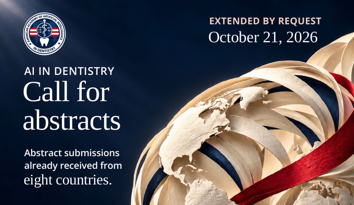
2027 Inaugural Annual Meeting — Call for Abstracts. By popular demand, the submission deadline has been extended to Wednesday, October 21, 2026. Abstracts have already arrived from eight countries — the United States, the United Kingdom, India, Canada, France, Italy, Egypt, and the Republic of Korea — a tremendous response, and we warmly welcome more. We will do our best to accommodate as many submissions as possible, provided they meet the requirements. Please send your abstract in the Academy’s structured format — Background, Methods, Results, and Conclusion — to abstracts@aaai-d.org by October 21. Acceptance notifications are scheduled for November 15, 2026.
New on the Education Hub: A White Paper, Regulatory Briefs, and a Learning Path for Patients
October brought the Academy’s largest publications push since its launch. The Academy has published a new white paper, AI Agents in the Dental Practice: A HIPAA Compliance Guide, in a new White Papers section of the website — free to read, with no login required. Written for practicing dentists, the guide sets out a practical framework for evaluating AI agents against HIPAA’s requirements, with worked examples from everyday dental workflows; it joins the Academy’s DSI Framework report in the section.
A new Regulatory Briefs section, also under Education, will track regulatory developments that matter to dental practice. Its first brief, “Two FDA Records in AI Worth Noting for Dentistry,” examines the FDA’s August 2026 discussion paper on generative and agentic AI and a 2025 adverse-event report involving a dental AI product — and what the two records show, together, about how AI oversight is taking shape.
And the Education hub itself has been rebuilt around a simple idea: patients deserve the same clarity about dental AI as clinicians. A new nine-step learning path explains, in plain language, what dental AI is, how it is reviewed and cleared, and what patients can ask their own dentists — alongside the Academy’s professional resources, Regulatory Briefs, and Research Highlights.
Los Angeles 2027: The Preliminary Program Arrives With Registration
The preliminary program for the Academy’s Inaugural Annual Meeting will be released very soon — and its release will coincide with the reopening of membership registrations and meeting registrations. The Academy’s membership and registration pages reopen soon.
The meeting takes place on Friday, July 9, 2027, in the Grand Horizon Room at UCLA Covel Commons in Los Angeles. The program will be published on the Academy’s website and shared with members as soon as it is released.
The Academy in Barcelona
On September 30, the Academy’s president, Dr. Owais Farooqi, gave an invited lecture at UIC Barcelona (Universitat Internacional de Catalunya), speaking to students and faculty on the state of AI in dentistry — its clinical applications, the evidence behind it and its limits, and the place of human judgment. Students’ questions were perceptive, including a debate over whether AI will improve or degrade the patient experience. While in Barcelona, Dr. Farooqi also met with UIC Barcelona faculty, including Prof. Miguel Roig Cayón, to discuss future collaboration between UIC and the Academy in AI education.
The next day, he opened the ‘Where we are today’ session of the AI in Dentistry panel at the starlynr® International Forum 2026, moderated by Ziad Matar, before a highly engaged audience of students and faculty. The Academy thanks Dr. Fady Yaacoub and the Starlynr team for the invitation.
One name in the next item will be familiar: Prof. Roig, of UIC Barcelona, is among the speakers recently confirmed for Los Angeles.
Recently Confirmed for Los Angeles 2027
The Academy continues to confirm speakers for its Inaugural Annual Meeting. Among those recently confirmed:
 Prof. Reinhilde Jacobs is a full professor at KU Leuven in Belgium, where she heads the OMFS-IMPATH research group and the university’s DentoMaxilloFacial Imaging Center; she also serves as Secretary General of the International Association of DentoMaxilloFacial Radiology. She will speak on the path from patient-specific modelling to intelligent treatment planning.
 Dr. Niklas Virta, chief executive of Aqua Dental — one of Sweden’s largest dental groups, with 37 clinics across the country — will report on what worked, and what did not, when his organization put AI solutions to work at scale.
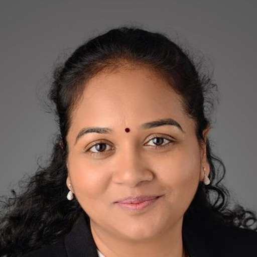 Dr. Aarthi Shanmugavel, ADA Standards Program Director at the American Dental Association in Chicago, will speak on the AI standards shaping dentistry’s future.
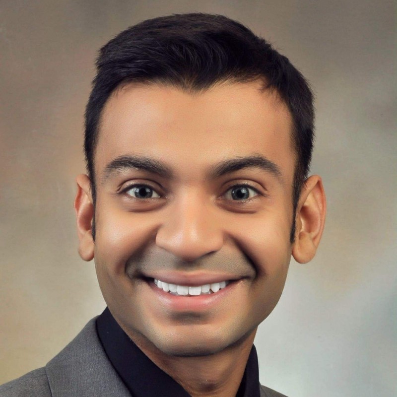 Dr. Jay S. Patel, Assistant Professor of Health Informatics and Dentistry at Temple University’s Kornberg School of Dentistry in Philadelphia, will speak on identifying high-risk patients in the dental record before the first symptom appears.
 Prof. Miguel Roig, Full Professor of Restorative Dentistry at UIC Barcelona in Spain, will speak on AI in restorative care and the move toward intelligent, precision dentistry.
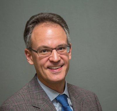 Prof. Michael Bornstein, professor and chair of the Department of Oral Health & Medicine at the University Center for Dental Medicine Basel, University of Basel in Switzerland, and Editor-in-Chief of Dentomaxillofacial Radiology, the journal of the International Association of DentoMaxilloFacial Radiology, will ask whether the AI-augmented dento-maxillofacial radiologist is utopia or dystopia.
The full preliminary program will be released soon, together with the reopening of membership and meeting registrations.
Sources
1. Nalbantoglu et al., Biruni University, Istanbul. AI-assisted caries detection on panoramic radiographs (4,000 first permanent molars). BMC Oral Health. September 28, 2026. DOI: 10.1186/s12903-026-09998-6.
2. Salman & Bishtawi, An-Najah National University. Multimodal AI grading of periodontal bone-resorption severity from panoramic radiographs and structured patient data. BMC Medical Imaging. October 13, 2026. DOI: 10.1186/s12880-026-02928-6.
3. Zhang et al., Lanzhou University. Artificial intelligence for detection of periodontitis on radiographic images: a systematic review and diagnostic test accuracy meta-analysis. Journal of Dentistry. September 17, 2026. DOI: 10.1016/j.jdent.2026.107054.
4. Bader. When AI Gives Dental Advice: Managing Patient Risk in a New Diagnostic Landscape. British Dental Journal 241, 450–453. October 9, 2026.
5. Li, Bao et al., Sun Yat-sen University, Guangzhou. Construction of a CNN model based on lateral cephalometric imaging features and its application in surgery-first treatment decision-making for skeletal Class III malocclusion. BMC Oral Health. September 25, 2026. DOI: 10.1186/s12903-026-09974-0.
6. Glick, Al-Hatem, Angelov & Chang, UTHealth Houston School of Dentistry. Presentation of AI-based decision support influences clinician decision making. BMC Medical Informatics and Decision Making. October 7, 2026. DOI: 10.1186/s12911-026-03871-w.
7. UofL News. Smart Smiles: How UofL Is Changing Dental Care With Cardinal Intelligence. University of Louisville School of Dentistry. October 2026.
8. İnönü University, Türkiye. Deep-learning detection of impacted maxillary canines on panoramic radiographs, with CBCT as reference standard. Diagnostics. September 29, 2026.
9. Şimşek et al., Mersin University. AI pulp-stone detection on panoramic radiographs (YOLOv8/YOLO11 benchmark; graph-neural-network hybrid). Scientific Reports. September 28, 2026.
10. Elçi et al., Necmettin Erbakan University. AI-assisted evaluation of midpalatal suture maturation using CBCT: a YOLOv11 study. BMC Medical Imaging. October 6, 2026. DOI: 10.1186/s12880-026-02886-z.
11. University of Bari “Aldo Moro” and University of Campania “Luigi Vanvitelli”, Italy. Artificial intelligence-based evaluation of oral potentially malignant disorders using photographic and clinical data. Dentistry Journal. October 10, 2026.
12. Zhang et al., University of Otago, Nanjing University, and Sichuan University. Deep learning-based automated detection and geometric severity assessment of open gingival embrasures in intraoral photographs: a multicentre study. Journal of Dentistry. September 17, 2026. DOI: 10.1016/j.jdent.2026.107055.
13. Bayrakdar et al. CRANIOSEG: Deep learning–based automated segmentation of craniomaxillofacial anatomical structures on cone-beam computed tomography. Dentomaxillofacial Radiology. September 15, 2026. DOI: 10.1093/dmfr/twag065.
14. Wang et al. Safety, accuracy, information quality, readability, and supportive communication of LLM-based chatbots in responses to researcher-developed consumer-oriented prompts on recurrent aphthous stomatitis. Frontiers in Oral Health. September 30, 2026. DOI: 10.3389/froh.2026.1968034.
15. Dawood et al., OMFS-IMPATH Research Group, KU Leuven. A novel AI-assisted 3D hybrid soft and hard tissue model for peri-implant diagnosis and treatment planning: a proof-of-concept study of immediate implant planning in the anterior maxilla. Journal of Dentistry. October 2, 2026. DOI: 10.1016/j.jdent.2026.107087.
16. Pagonis. Teaching AI-informed diagnostic reasoning in dental education: a conceptual framework. Journal of Dental Education. September 21, 2026. DOI: 10.1002/jdd.70389.
17. Li, Chen & Liao, Nankai University (Tianjin Stomatological Hospital). A closed-loop AI virtual simulation platform for preclinical fiber post-core restoration: two-year prospective evaluation of learning outcomes and student perceptions. BMC Medical Education. October 1, 2026. DOI: 10.1186/s12909-026-10510-5.
18. Chen, Sharma & An, Griffith University. Evaluating the effectiveness of artificial intelligence (AI)-supported removable partial denture design tools in dental education. Journal of Dental Education. October 6, 2026. DOI: 10.1002/jdd.70395.
19. Ozturk et al., Biruni University, Istanbul. Development and internal validation of a deep learning system for criterion-specific assessment of preclinical dental soap carvings: a multi-rater study. BMC Medical Education. October 7, 2026. DOI: 10.1186/s12909-026-10554-7.
20. Ribeiro et al., Suprema – Faculty of Medical and Health Sciences of Juiz de Fora, Brazil. Artificial intelligence in dental examinations: performance analysis of ChatGPT, Gemini, and undergraduate students. European Journal of Dental Education. September 30, 2026. DOI: 10.1111/eje.70319.
The AAAI-D Brief is published by the American Academy of Artificial Intelligence in Dentistry® for educational purposes. It summarizes published research and reporting and does not constitute clinical, legal, or regulatory advice. References to specific products, companies, or services are for informational purposes only and do not constitute endorsement, recommendation, or certification by AAAI-D.